Use of Endoscopic Diagnostic and Surgical Techniques at Nairi Medical Center
Endoscopy is a method of examining internal organs using a specialized optical instrument, the endoscope. Gastrointestinal endoscopy is considered one of the primary and most informative diagnostic tools that does not require open surgery and is widely used in clinical practice. During the procedure, an endoscope equipped with a distal optical system and a video camera provides real-time visualization of different segments of the gastrointestinal tract. An experienced specialist can identify pathological changes at a very early stage, which allows timely treatment planning.
Who requires endoscopic evaluation
Endoscopic examination may be performed based on patient complaints or as part of preventive screening. According to current clinical guidelines, upper GI endoscopy (EGD) is recommended at least once a year, and colonoscopy once every three years. After the age of 40, endoscopic evaluation becomes part of routine surveillance. This approach allows early detection of both benign and malignant lesions of the digestive tract, often at a stage when organ-preserving, minimally invasive endoscopic removal is still possible. Early intervention substantially improves the likelihood of cure compared to advanced disease.
Modern technologies have largely eliminated absolute contraindications to endoscopy. Gastroscopy can be performed under general anesthesia under continuous anesthesiology monitoring, ensuring the patient's comfort during and after the procedure. The examinations are relatively short, although the exact duration depends on individual clinical factors.
New methods in endoscopy
POEM (Peroral Endoscopic Myotomy).
Peroral endoscopic myotomy offers the advantage of treating esophageal outflow obstruction without abdominal incisions and without the uncontrolled risk of esophageal perforation associated with balloon dilation. This technique allows controlled myotomy (muscle fiber division) along a longer segment of the esophagus. POEM has demonstrated favorable safety in terms of infectious risk, hemodynamic stability, and respiratory and gastrointestinal tolerance. No major complications such as mediastinitis or peritonitis were reported.
Endoscopic ultrasound (EUS).
Endoscopic ultrasound enables high-resolution ultrasound imaging from within the gastrointestinal lumen, in close proximity to the structures being examined. This method allows detailed assessment of internal organs and identification of abnormalities that are not visible on conventional transabdominal ultrasound. The examination is performed using an endoscope that incorporates both an optical system and a miniaturized ultrasound transducer.
Unlike standard ultrasound, where signal quality decreases with depth and certain regions remain inaccessible, EUS can directly assess the esophagus, stomach, duodenum, colon and rectum from inside the lumen. It also provides access to adjacent structures in the abdominal cavity — including the pancreas, biliary tree, gallbladder and liver — and, via the esophagus, mediastinal structures in the chest. EUS can also evaluate lymph nodes in the thoracic and abdominal cavities.
Elastography during EUS
Sonoelastography performed during EUS provides high sensitivity, specificity and diagnostic accuracy in differentiating benign and malignant lymph node changes. Elastography complements standard EUS imaging modes and improves targeting for fine-needle aspiration biopsy in cases of multiple lymph node involvement.
Main clinical indications for EUS include:
- Evaluation of benign (polyps, submucosal lesions, cysts, extrinsic compression) and malignant lesions of the esophageal, gastric and duodenal wall. EUS can determine the layer of origin, depth of invasion and presence of regional lymphadenopathy.
- Diagnosis of pancreatic disease (acute and chronic pancreatitis, cysts, neoplasms, intraductal stones).
- Assessment of gallbladder and biliary tract pathology, particularly in distal bile ducts and the ampullary region, which are difficult to visualize by other techniques.
- Evaluation of mediastinal masses.
- Assessment of variceal dilatation of the esophageal and gastric veins in portal hypertension, and planning of bleeding prevention and management.
- Identification of deep collateral venous pathways.
EUS is particularly valuable in the early diagnosis of:
- Submucosal tumors of the esophagus, stomach, duodenum and colon (e.g. fibroma, lipoma, leiomyoma),
- Carcinoma of the esophagus, stomach, duodenum and colon,
- Esophageal varices,
- Peptic ulcer disease and structural deformities,
- Biliary obstruction, including choledocholithiasis,
- Ductal lesions and cystic changes of the pancreas.
At Nairi Medical Center, endoscopic procedures are performed using the PENTAX Hi-Line HD+ digital videoendoscopy platform (Japan). This HD+ system offers image clarity superior to standard endoscopy systems. Its advanced image enhancement technology allows detailed examination of the mucosa and detection of minimal pathological changes, including precancerous lesions and early-stage malignancies. The i-scan function improves visualization of mucosal architecture and facilitates rapid lesion detection. Virtual chromoendoscopy makes it possible to characterize lesions without chemical dyes, reducing examination time.
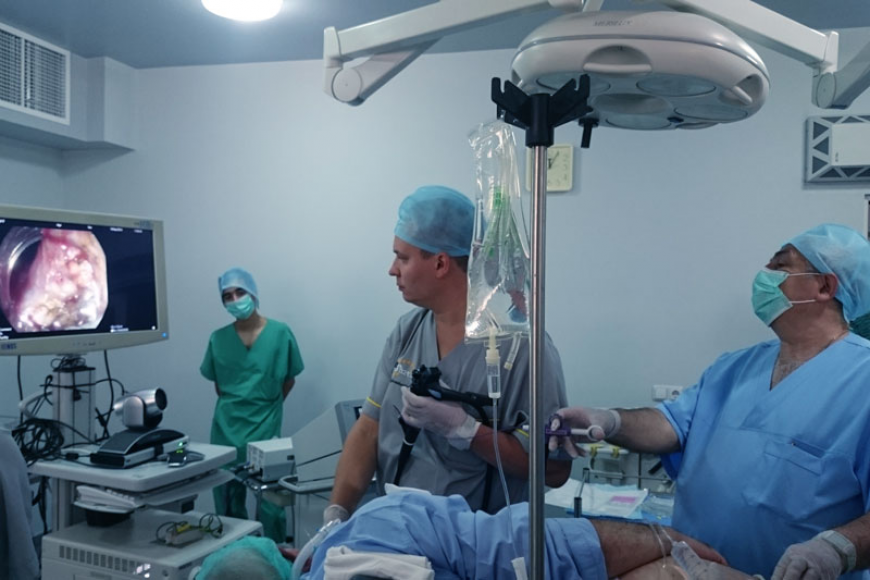
On September 10, 2016, at Nairi Medical Center, a series of complex therapeutic and diagnostic endoscopic interventions and EUS procedures were performed for the first time in Armenia using expert-class equipment. The invited specialist, Dr. Aleksandr Smirnov, PhD (Saint Petersburg), Head of the Department of Endoscopic and Emergency Surgery and lecturer at the Pavlov State Medical University, conducted live procedures and a master class. The interventions were broadcast in real time from the operating room to the conference hall for observing physicians.
The clinical cases included:
- Peroral endoscopic myotomy (POEM) for esophageal achalasia.
- EUS with biopsy in patients with suspected neoplasm of the pancreatic head.
- EUS with biopsy in cases of Barrett’s esophagus.
- Endoscopic mucosal and submucosal dissection to remove colonic lesions.
- Diagnostic EUS in a patient with a gastric lesion.
Most patients were discharged the same day, and the rest on the following day.
Such interventions require high-end technical infrastructure, qualified anesthesiology support and careful postoperative supervision. A key advantage is rapid recovery and early return to normal activity with minimal impact on quality of life.
Endoscopic ultrasound has a distinct advantage over conventional ultrasound, CT and standard endoscopy. It enables early detection of clinically significant pathology in otherwise inaccessible anatomical regions and permits precise, ultrasound-guided targeted biopsy. This substantially improves both diagnostic accuracy and therapeutic decision-making in diseases of the gastrointestinal tract.